
Retinal Detachment
What is Retinal Detachment?
A retinal detachment is a serious condition whereby the retina detaches from its normal position on the back wall of the eye. This results in blindness in that portion of the vision. In many cases, the problem can be corrected with surgery.
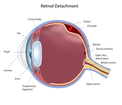
The retina is a very thin layer of specialized nerve cells that receive light from the outside world and convert it to electrical impulses that are sent to the brain. If the eye were a camera, the retina would be the film. Trauma and various disease processes can cause a retinal detachment, with many occurring in people with myopia (nearsightedness).
A primary cause of retinal detachment is the formation of a hole or tear in the retina which allows fluid to pass behind the retina, elevating it from its normal position. For example, retinal holes or tears may occur from pulling forces of the vitreous, as it liquefies and contracts with age. Vitreous is the clear gel-like substance that fills the eye. Small condensations of the vitreous gel first appear as “floaters”, seen as floating webs, strands or dust-like specks in our vision. Generally, “floaters” are not a major concern, only a sign that some change in the vitreous has taken place.
However, the sudden occurrence of new floaters may be the first symptom of a retinal hole or tear which could lead to a retinal detachment. In addition, the occurrence of “flashes”, appearing as sudden flashes of light in any part of our vision, is potentially a more serious sign that the vitreous gel is pulling on a portion of the retina. This more intense pulling could result in the formation of a tear or hole in the retina at that location. The occurrence of “floaters” or “flashes” should be checked by a qualified eye care specialist as soon as possible, along with any other noticeable changes in your vision.
When a full retinal detachment has occurred a common surgical repair called a “scleral buckling procedure” is sometimes prescribed. It involves the use of a small silicone band placed around the eye to reposition the retina back in its normal location. Additionally, drainage of the fluid beneath the retina and sealing of the tear with heat, freezing or laser may be performed. As with any surgical procedure, side effects and complications may occur. If caught early, symptomatic retinal holes and tears often can be prophylactically treated with an in-office laser treatment or outpatient freezing procedure which “welds” the retinal hole/tear closed. With these advanced techniques, many retinal detachments can be prevented if caught at an early stage. This is why it is imperative to immediately report any new floaters/flashes or sudden visual changes to your Eye Care Specialist.
How a Detachment Occurs
- The vitreous may pull on the retina
- The tugging vitreous can cause the retina to tear
- Fluid can pass through the tear and behind the retina, lifting it.
- As more fluid goes behind the retina, it detaches further.
The Treatment
One of the most common and successful treatments for retinal detachment is called scleral buckling surgery. A small silicone band is placed around the eye which causes the retina to reattach to its original position. Tears in the retina may be sealed with a laser or by other means.

The doctors at Kovach Eye Institute have either authored or reviewed and approved this content.
Page Updated: